Flow and transport in the central nervous system
The cerebrospinal fluid (CSF) is a colorless fluid that bathes the external surfaces of the brain and the spinal cord. At normal body temperatures, the CSF is an incompressible Newtonian fluid with constant density and kinematic viscosity similar to those of water. The CSF exhibits a fast oscillation driven by the pressure differences induced by the cardiac and respiratory cycles and a much slower Lagrangian mean motion. The associateory motd transport rate is key for maintaining the electrolytic environment, transporting hormones, circulating nutrients and chemicals filtered from the blood, and removing waste products from the cell metabolism of the brain and the CNS. Deregulated CSF circulation has been reasoned to play a role in the development of various neurological disorders, including normal pressure hydrocephalus, syringomyelia and Alzheimer’s disease, for example. Our work uses a combination of theoretical and numerical techniques informed by in-vitro experiments and in-vivo magnetic-resonance imaging to provide fundamental understanding and accurate quantification of different key flow and transport processes involving CSF. The investigation is supported by the National Institute of Neurological Disorders and Stroke through contract No. 1R01NS120343-01 and by the National Science Foundation through grant number 1853954.

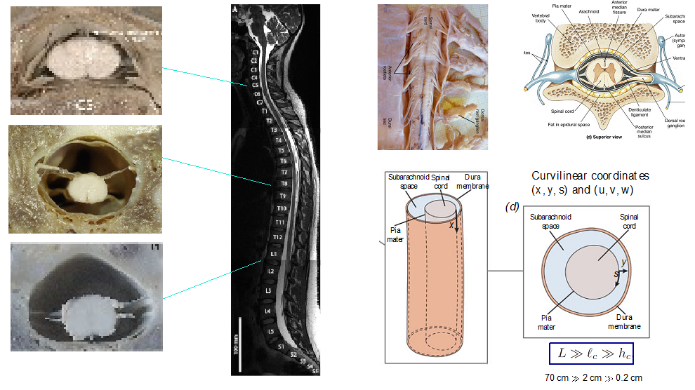
The spinal canal anatomy, including a general view as well as detailed images of different key features.


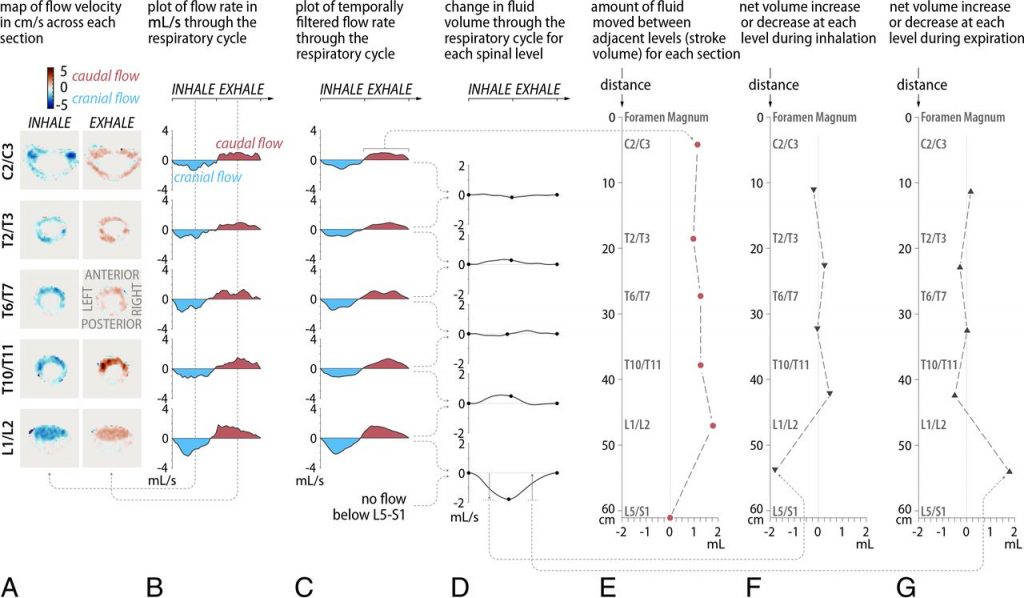
In-vivo experimental investigation of respiration-driven flow in the spinal canal. The upper panel show a schematic overview of the MR imaging setup. The lower panels show sample MR results, including A, Map of flow velocity in centimeters per second across each section at mid-inhalation (leftcolumn) and mid-expiration (right column). B, Plot of the flow rate in milliliters per second across each section through the respiratory cycle. C, Plot of temporally filtered flow rate. D, Change in fluid volume through the respiratory cycle with respect to the start of inhalation for each spinal level. E, Amount of fluid moved between adjacent levels (stroke volume) for each section. F, Net volume increase or decrease at each level during inhalation. G, Net volume increase or decrease during expiration.

Analysis of peristaltic motion in non-axisymmetric annular tubes motivated by observations of flow along periarterial spaces in the brain and spinal cord.
Related publications
-
- On the bulk motion of the cerebrospinal fluid in the spinal canal
A. L. Sánchez, C. Martínez-Bazán, C. Gutiérrez-Montes, E. Criado-Hidalgo, G. Pawlak, W. Bradley, V. Haughton, J. C. Lasheras, , J. Fluid Mech, 841 203–227 (2018). [DOI] - Lubrication analysis of peristaltic motion in non-axysimmetric annular tubes
W. Coenen, X. Zhang, A. L. Sánchez, J. Fluid Mech., 921 R2 (2021). [DOI] - Effect of normal breathing on the movement of CSF in the spinal subarachnoid space
C. Gutiérrez-Montes, W. Coenen, M. Vidorreta, S. Sincomb, C. Martínez-Bazán, A. L. Sánchez, and V. Haughton, Am. J. Neuroradiol., 43 1369–1374 (2022). [DOI] - The directional flow generated by peristalsis in perivascular networks – theoretical and numerical reduced-order descriptions
I. Gjerde, M. E. Rognes, A. L. Sánchez, J. Appl. Phys., 134, 174701 (2023) [DOI] - An analytic model for the flow induced in syringomyelia cavities
G. L. Nozaleda, J. Alaminos-Quesada, W. Coenen, V. Haughton, A. L. Sánchez, J. Fluid Mech., 978, A22 (2024) [DOI]
- On the bulk motion of the cerebrospinal fluid in the spinal canal
Development of patient-specific mathematical models for intrathecal drug delivery
The treatment of a number of central-nervous-system (CNS) pathologies, including some cancers of the CNS, as well as the management of severe chronic and post-operative pain involve in some cases the direct injection of the medication into the CSF in the intrathecal space of the spinal canal that surrounds the spinal cord. This procedure, used since the early 1980s, is often referred to as intrathecal drug delivery (ITDD). The standard ITDD protocol consists of placing a small catheter along the spine in the SAS of the lumbar region to continuously pump the drug or to release a finite dose at selected times. Sometimes ITDD is used to deliver the drug to sites along the spinal cord close to the location of injection, while in other cases the medication is delivered to distant target sites in the brain. Although ITDD is currently used with satisfactory results, the drug dispersion rate is rather unpredictable and exhibits dependences that are not thoroughly understood. Our work, combining multiscale analyses with MRI measurements of the flow, is motivated by the existing need to develop a methodology capable of accurately predicting the dispersion of the drug along the spinal canal for the specific anatomy and physiological conditions of the individual patient as well as for the molecular characteristics and injection rate of the drug. The work is supported by the National Institute of Neurological Disorders and Stroke through contract No. 1R01NS120343-01 and by the National Science Foundation through grant number 1853954.

Sagittal T2-weighted MR image of the spine for a subject in the supine position (A), diagram of the sagittal images (B) and axial images (C), diagrams of the images at selected locations along the spine (D), width of the SSAS projected onto a 2D plane obtained by unrolling the space (E), and 2D map of the width of the SSAS after converting the length and circumference to dimensionless units (F). The right-hand-side plot shows corresponding streamlines and direction for bulk (mean Lagrangian) flow of the CSF in the projected onto the 2D dimensionless plane with anterior midline SSAS at s=0 and the posterior one at s=0.5.
Related publications
-
-
- On the dispersion of a drug delivered intrathecally in the spinal canal
J. J. Lawrence, W. Coenen, A. L. Sánchez, G. Pawlak, C. Martínez-Bazán, V. Haughton, J. C. Lasheras, ,J. Fluid Mech., 861 679–720 (2019) . [DOI] - Subject-specific studies of CSF bulk flow patterns in the spinal canal: implications to the dispersion of solute particles in intrathecal drug delivery
W. Coenen, C. Gutiérrez-Montes, S. Sincomb, E. Criado-Hidalgo, K. Wei, K. King, V. Haughton, C. Martínez-Bazán, A. L. Sánchez, and J. C. Lasheras, , Am. J. Neuroradiol., 40 1242–1249 (2019). [DOI] - Modelling and direct numerical simulation of flow and solute dispersion in the spinal subarachnoid space
C. Gutiérrez-Montes, W. Coenen, J.J. Lawrence, C. Martínez-Bazán, A. L. Sánchez, Applied Mathematical Modelling, 94 516–533 (2021). [DOI] - Buoyancy-modulated Lagrangian drift in wavy-walled vertical channels as a model problem to understand drug dispersion in the spinal canal
J. Alaminos-Quesada, W. Coenen, C. Gutiérrez-Montes, A. L. Sánchez, J. Fluid Mech., 949, A26 (2022) [DOI] - In vitro characterization of solute transport in the spinal canal
F. Moral-Pulido, J. I. Jiménez-González, C. Gutiérrez-Montes, W. Coenen, A. L. Sánchez, C. Martínez-Bazán, Phys. Fluids, 35, 051905 (2023) [DOI] - Effects of buoyancy on the dispersion of drugs released intrathecally in the spinal canal
J. Alaminos-Quesada, C. Gutiérrez-Montes, W. Coenen, A. L. Sánchez, J. Fluid Mech., 985, A20 (2024) [DOI]
- On the dispersion of a drug delivered intrathecally in the spinal canal
-
Non-invasive evaluation of intracranial pressure
The intracranial pressure (ICP) plays a central role in the development of many neurological diseases. In general, monitoring the mean ICP, its temporal fluctuations and spatial variations requires the insertion of pressure sensors, an invasive procedure with considerable risk factors. Temporal intracranial pressure fluctuations drive the wave-like pulsatile motion of cerebrospinal fluid (CSF) along the compliant spinal canal. Similarly, the so-called transmantle pressure, the instantaneous spatial pressure difference between the lateral ventricles and the cranial subarachnoid space, drives cyclic CSF flow in and out of the ventricles through the cerebral aqueduct. Therefore, systematically derived simplified models relating the ICP temporal and spatial fluctuations with the resulting CSF flow rate can be useful in enabling indirect evaluations of the former from non-invasive magnetic resonance imaging (MRI) measurements of the latter. The work is supported by the National Institute of Neurological Disorders and Stroke through contract No. 1R01NS120343-01 and by the National Science Foundation through grant number 1853954.

A model for the flow in the cerebral aqueduct: (a) Schematic views of the cranial cavity and (b) the cerebral ventricular system. Anatomic magnetic resonance images are used to obtain (c) a smoothed surface mesh of the cerebral aqueduct of a healthy subject, which was used for (d) the simplified illustration highlighting the different flow regions and their reduced mathematical description.

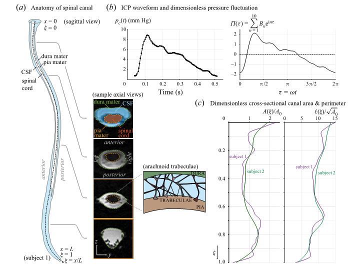
(a) Main anatomical features of the spinal canal; (b) typical ICP wave form (c) dimensionless canal cross-sectional area and perimeter for two subjects.
Related publications
-
-
-
- A model for the oscillatory flow in the cerebral aqueduct
S. Sincomb, W. Coenen, A. L. Sánchez, J. C. Lasheras ,, J. Fluid Mech., 899 R1 (2020). [DOI] - Transmantle pressure computed from MR measurements of aqueduct flow and dimensions
S. Sincomb, W. Coenen, E. Criado-Hidalgo, K. Wei, K. King, M. Borzage, V. Haughton, A. L. Sánchez, and J. C. Lasheras, Am. J. Neuroradiol., 42 1815–1821 (2021). [DOI] - A one-dimensional model for the pulsating flow of cerebrospinal fluid in the spinal canal
S. Sincomb, W. Coenen, C. Gutiérrez-Montes, C. Martínez-Bazán, V. Haughton, A. L. Sánchez, J. Fluid Mech., 939 A26 (2022) . [DOI] - An in vitro experimental investigation of oscillatory flow in the cerebral aqueduct
S. Sincomb, F. Moral-Pulido, O. Campos, C. Martínez-Baz\'{a}n, V. Haughton, A. L. Sánchez, Eur. J. Mech. B/Fluids., 105, 180-191 (2024) [DOI]
- A model for the oscillatory flow in the cerebral aqueduct
-
-